Mammography is a specialized type of low-dose X-ray imaging that healthcare practitioners use to help them look for abnormalities in breast tissue.
Radiologists routinely use mammograms in two important ways, both related to breast cancer:
- Screening mammograms are performed at routine intervals to look for signs of breast cancer in women who don’t have any symptoms.
- Diagnostic mammograms look more closely at abnormalities in the breast tissue of women who have identified symptoms consistent with breast cancer, or who have suspicious findings on a screening mammogram.
Traditional mammograms capture images of breast tissue in two dimensions (2D). In recent years, the more advanced 3D mammography has become increasingly common at medical facilities due to its advanced capabilities and improved patient outcomes.
What Is 3D Mammography?
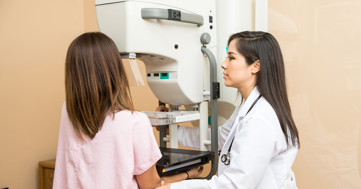
Also referred to as breast tomosynthesis, 3D mammography can be used for both diagnostic and screening purposes. During the imaging session, each breast is compressed once, and an imaging device takes many low-dose X-ray images as it moves in an arc over the breast.
This creates a series of 200-300 images, instead of the four yielded by a traditional 2D mammogram. These hundreds of images are then stitched together by a computer into a three-dimensional image, allowing radiologists to study glandular breast tissue in greater detail than they could with 2D mammograms alone.
How Are 3D Mammograms Different From 2D?
While the patient’s experience of a 3D mammogram procedure is about the same as that of a 2D mammogram, many practitioners believe that 3D imaging yields better diagnostic results.
2D mammograms only capture images of breast tissue from the top and side of the breast. These two-dimensional images allow overlapping tissues to block the low-dose X-rays, potentially hiding tumors.
3D mammograms empower radiologists to study the breasts from many different angles, effectively looking “behind” any overlapping tissues that might otherwise block their view.
Better Results and Fewer Follow-Ups
With their ability to capture tissue from many more angles, 3D mammograms are reducing the need for follow-up imaging. With 2D mammograms, patients sometimes have to go for follow-up tests to further study any abnormalities revealed in the initial mammogram. Research suggests patient callbacks have decreased 15-30% in centers where 3D mammograms have been implemented.
3D mammography may also be better suited for women with dense breast tissue. The breast is made up of milk ducts, milk glands, supportive tissue, and fatty tissue. Some women naturally have more supportive, dense breast tissue than others. In standard mammography, it can be challenging to detect signs of breast cancer in areas of density. With 3D mammography, doctors can see past these areas of density for better screening.
Most importantly, research suggests 3D mammography could detect more breast cancers in the early stages of their development. Studies are underway to look more closely at clinical outcomes between 3D and 2D mammography. With that being said, any chance that breast cancer could be detected earlier is important, as treatment outcomes are best in the disease’s earliest stages. In many cases, mammography can detect cancer before a lump can even be felt.
If you’re due for a mammogram, request an appointment with Outpatient Imaging Culpepper online or by calling (540) 321-3190. You can also learn more about what to expect from a 3D mammogram.